How a new embryo model can tell us how life starts
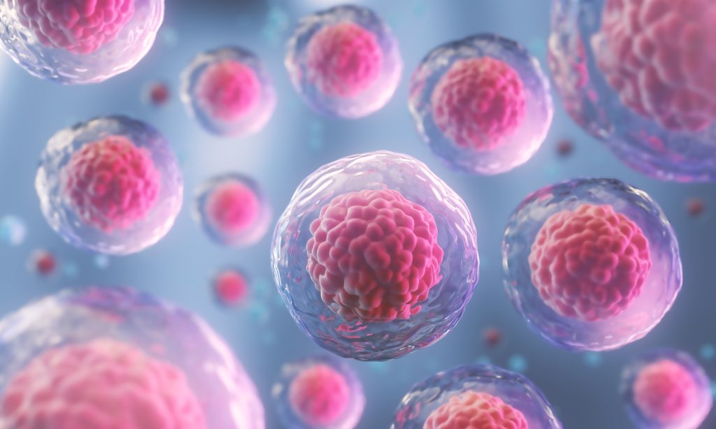
Human embryogenesis, or the development of a human embryo, is a complex and long-studied topic in developmental biology. Studying embryogenesis provides insight into how certain diseases develop and possible solutions. The process comprises many cycles of cell division and tissue formation, which are difficult to study due to their intricate nature and the ethics involved in research. Stem cells have been used in recent years to mimic the development of human embryos. However, these models have flaws that obstruct our understanding of true embryogenesis. This article has crafted an alternative model that avoids these flaws and provides the scientific community with a method to study early embryo development and blood formation.
A New Model for Embryos
There are few effective models for studying the development of human embryos. Existing models typically lack extra-embryonic tissues, the development of which is still largely unstudied, and are difficult to maintain. The investigators have created a novel model, termed ‘heX-embryoid,’ to support researchers studying the extra-embryonic niche and yolk sac hematopoiesis, both vital to understanding embryogenesis.
To develop the heX-embryoid model, the investigators used human-induced pluripotent stem cells (hiPS cells). These mature cells have been reprogrammed into a special type of cell, pluripotent cells, that can give rise to any cell type in the body. This reprogramming is done so that the cells mimic the state of embryonic cells, which can then be manipulated and studied further. In this article, the researchers show how their model was developed and accommodates a myriad of potential research that can tell us about embryogenesis and possible solutions for diseases.
What They Found
Most researchers will rely on model organisms to study their topic, but current models are insufficient for labs studying amniogenesis – the development of the amniotic cavity. After the heX-embryoid was generated through the genetic addition of the human gene, GATA6, rigorous testing was undertaken to determine limitations and advantages, including the ability to form the amnion. To test this, they identified gene markers or genes that express in human amnion. They found that the ISL1, TFAP2A, and GATA3markers were present in the heX-embryoid, indicating it had undergone amniogenesis, a process otherwise difficult to replicate in a lab.
Also of great importance to developing embryos is the yolk sac, which provides nutrition to the organism and kicks off early hematopoiesis, or the production of blood cells. Hematopoiesis begins within the tissues of the embryo’s yolk sac, and understanding the molecular mechanisms of this process is vital to understanding embryogenesis as a whole. Structural image analysis of the heX-embryoids found that they were morphologically similar to developing human embryos (Fig 4k from published paper). This similarity was confirmed by looking at how the tissues are arranged based on which gene markers are present in each tissue.
These features of the heX-embryoid could allow other labs to study drug testing and developmental toxicology without technical or ethical hindrances.
Not so fast
Every model organism has its drawbacks, and this is also true here. While the heX-embryoid offers significant morphological similarities to human embryos previously unavailable, it is still a model. Results from experiments tested on the heX-embryoids may closely resemble the expected results from real embryos, but they are not perfect predictors. The importance of this approach is that results tested on this model can serve as a guide for further research.
Why does it matter?
The heX-embryoid offers labs a model for human embryogenesis that sidesteps typical technical and ethical hurdles. Additionally, this model can be used for live imaging and requires fewer materials than alternatives. This tool could enable breakthrough studies on developmental biology and modelling diseases.
Take home messages
1. heX-embryoid is a new model system for human embryo development.
2. heX-embryoid can be made easily available to labs researching developmental biology.
3. This model will provide insight into the development of human embryos, although it cannot perfectly replicate real human development.
Guest author: Gabrielle Dubansky, M.S.
This article was written as part of a series of ‘journal club’ summaries for Scientific Writers Ltd., and is based on the following publication:
Title: Modelling post-implantation human development to yolk sac blood emergence
First Author: Hislop J, et al.
Journal: Nature
Date online: 13 December 2023