A Place for Everything, and Everything in its Place
Parkinson’s Disease (PD) features identifiable motor symptoms including tremor, rigidity and slowness of movement, often accompanied by balance issues and freezing of gait. In PD patients, Streptococcus mutans (S mutans), a bacterium usually found in the mouth, colonises the gut. The misplaced presence of this particular resident is far from harmless and correlates with PD pathology via a defined signalling pathway.
Explaining Parkinson’s Disease
PD is characterised by selective loss of dopaminergic neurons and the alteration of a protein (aggregated α-synuclein) in a specific region of the brain called the substantia nigra pars compacta. Although PD may have a genetic component, the condition is considered idiopathic in most cases, indicating that environmental factors (eg, viruses, metals, air pollution, pesticides) play an important role in disease onset.¹
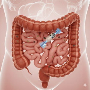
The gut’s microbiome is also influenced by environmental factors. The connection between gut microbiota and PD has been extensively explored, revealing that PD patients have altered bacterial gut profiles. Experiments in PD animal models show that faecal microbiota from patients with PD can promote PD-like symptoms in otherwise healthy animals. Until recently, the specific bacteria that contribute to PD development had not been identified. A 2022 metagenomic study revealed that the bacterium S mutans was enriched in the gut of PD patients.² This microbe is commonly found in the human mouth, so its conspicuous presence in the gut suggests a possible role in PD pathology. But how could the appearance of a bacterium in the gut bring on a complex neurodegenerative disorder?
Park et al (2025) set out to determine whether S mutans can, in fact, colonise the gut and affect PD pathology via microbial production of imidazole propionate (ImP).
The imidazole propionate link
S mutans can degrade the amino acid histidine, generating the molecule ImP. Once produced, ImP can travel through the bloodstream from the gut lumen to the brain and disrupt specific dopaminergic neurons, a hallmark characteristic of PD.
To investigate the link between ImP and PD, investigators inoculated germ-free mice with S mutans. Pasteurised samples of the bacteria (ie, dead S mutans) were used to inoculate mice in the control group. After the inoculation period, they found viable S mutans in the experimental animals’ faeces, but not in the control group. This finding confirmed that S mutans colonised the mouse gut. Researchers also demonstrated that S mutans colonisation provoked neurotoxicity in the animals’ midbrain, inducing PD-related changes: selective loss of dopaminergic neurons, astrogliosis, microgliosis and motor symptoms.
Next, investigators measured the level of ImP in the animals’ bloodstream and brain tissue. The metabolite was present in both the bloodstream and brain of animals exposed to live S mutans, but not in animals inoculated with pasteurised S mutans.
The molecular circuit, exposed
ImP alters insulin signalling in hepatocytes (liver cells) by activating the p38γ-mTORC1 pathway (also called mTORC1). This circuit is deeply connected with ageing and neurodegenerative diseases.
The researchers wanted to know if S mutans gut colonisation was sufficient to activate the mTORC1 pathway in the midbrain. To answer this question, they screened for the specific activation of the mTORC1 pathway using an immunofluorescence technique. This technique integrates microscopy, fluorescence, and specific antibody-mediated recognition of proteins to discern if a particular pathway is on or offin a given neuron.
They determined that gut S mutans led to mTORC1 activation specifically in dopaminergic neurons of the substantia nigra pars compacta, without affecting mTORC1 signalling in other areas of the brain. Please refer to Figure 3 of the manuscript for further details and a view of the fluorescent images that were generated, which show the results linking S mutans presence to mTORC1 activation in specific brain regions. This result underscores the key role played by ImP-induced neuronal toxicity in PD pathogenesis by activation of the mTORC1 pathway.
Finally, the investigators explored the possibility of reversing the effect of S mutans gut colonisation by turning off the mTORC1 pathway using rapamycin, which specifically inactivates mTORC1. As expected, rapamycin treatment did not affect S mutans gut colonisation of germ-free mice. It was also noted that rapamycin:
– Did not reduce the elevated levels of ImP in plasma and brain, induced by S mutans gut colonisation,
– Reversed the decrease in brain weight that was induced by S mutans,
– Effectively abolished the brain abnormalities provoked by S mutans gut colonisation,
– Prevented dopaminergic neurodegeneration, astrogliosis, and microgliosis in the midbrain, as well as other alterations, and
– Reversed the motor symptoms induced by S mutans colonisation.
Not so fast
Though these results certainly suggest a solid link between S mutans found in the gut and PD, the study findings need further investigation. First, longitudinal studies of human cohorts are required to firmly establish whether gut microbial production of ImP and the subsequent signalling events initiate PD pathologies.
Next, the observed astrogliosis and microgliosis do not fully prove neuroinflammation. Cytokine profiling and other well-established markers are needed to determine the existence of a broader neuroinflammatory response. Also, both the motor and non-motor consequences of ImP and S mutans gut colonisation require further exploration. Future studies should include a broader range of PD signs and symptoms (eg, depression, olfactory deficits, cognitive impairment, anxiety, constipation).
Research that examines the effects of ImP on non-dopaminergic brain regions is limited and additional studies in this area may shed light on PD and other brain pathologies.
Why does it matter?
This research uncovers a direct gut-brain link in PD, showing how a common oral bacterium, S mutans, colonises the gut, produces ImP, and triggers neurodegeneration. The results fill a key knowledge gap by identifying a brain-permeable microbial metabolite (ImP) as a cause for selective midbrain dopaminergic neurotoxicity.
Management of the ImP/mTORC1 pathway could represent a novel therapeutic target and opens the door to new strategies in PD diagnostics (eg, blood ImP/UrdA testing). The finding that rapamycin can reverse some effects of S mutans gut colonisation represents (with future study) a potential to develop a major therapeutic intervention for millions of patients with PD.
Taking a more global perspective, this work provides evidence for gut microbes as PD drivers, identifying targets that could lead to PD prevention via lifestyle³ and gut-focused interventions (eg, probiotics or faecal transplants). It supports broader visions for averting PD onset using environmental/microbiome strategies, such as identification of enabling biomarkers (ImP testing) and early intervention therapies.
Take home messages
1. The gut microbiome of PD patients shows elevated S mutans, producing ImP.
2. S mutans colonises the mouse gut, elevates systemic/brain ImP levels, and induces numerous hallmarks of PD.
3. ImP penetrates the brain and activates mTORC1 selectively in dopaminergic neurons, driving neurotoxicity.
4. Rapamycin blocks the neurotoxic effects of gut S mutans.
Guest author: Celina Galles, PhD
Reviewers: Barbara Fahmy, MS OTR, MPA; Allison Kirsop, PhD
This article was written as part of a series of ‘journal club’ summaries for Scientific Writers Ltd and is based on the following publication.
Title: Gut microbial production of imidazole propionate drives Parkinson’s pathologies
First Author: Hyunji Park
Journal:Nature Communications
Date online: 5 September 2025
Other References:
- Dorsey ER, Bloem BR. Parkinson disease is predominantly an environmental disease. J Parkinsons Dis. 2024;14(3):451 465. https://doi.org/10.3233/JPD-230357
- Wallen, Z.D., Demirkan, A., Twa, G. et al. Metagenomics of Parkinson’s disease implicates the gut microbiome in multiple disease mechanisms. Nat Commun 13, 6958 (2022). https://doi.org/10.1038/s41467-022-34667-x
- Trinh J, de Vries NM, Chan P, et al. The role of lifestyle interventions in symptom management and disease modification in Parkinson disease. Lancet Neurol. 2025;24(1):90 102. https://doi.org/10.1016/s1474-4422(25)00305-9