A new sensor based on tiny gold particles could help improve the understanding of lung and airway chemistry in people living with Cystic Fibrosis.

Cystic Fibrosis (CF) is a life-limiting genetic disease and the leading genetic cause of death among the Caucasian population; one in every 2500 children in the UK is born with CF. People with this disease experience chronic lung infections and inflammation, and understanding how the disease influences lung and airway chemistry could help develop new therapies and decisions on how best to personalise existing treatments.
What the researchers uncovered
Multi-disciplinary research led by Prof. Campbell’s group at the University of Edinburgh is making it easier to understand key aspects of airway chemistry. Sensors smaller than the width of a human hair that sit in the thin layer of liquid on the airway surface have been developed. These sensors are intended for personalised models of airways that are routinely grown in the lab from the cells of people with CF, to test how patients will respond to therapies.
This study highlights how these sensors, made from a microscale polystyrene core surrounded by gold nanoparticles, interact with laser light to generate a strong signal that is characteristic of the airway chemistry. The sensors sit on the airway surface and can report on the activity of the key mutated protein in people with CF. Importantly, it could provide a means of identifying when a patient will respond positively to a particular drug combination.
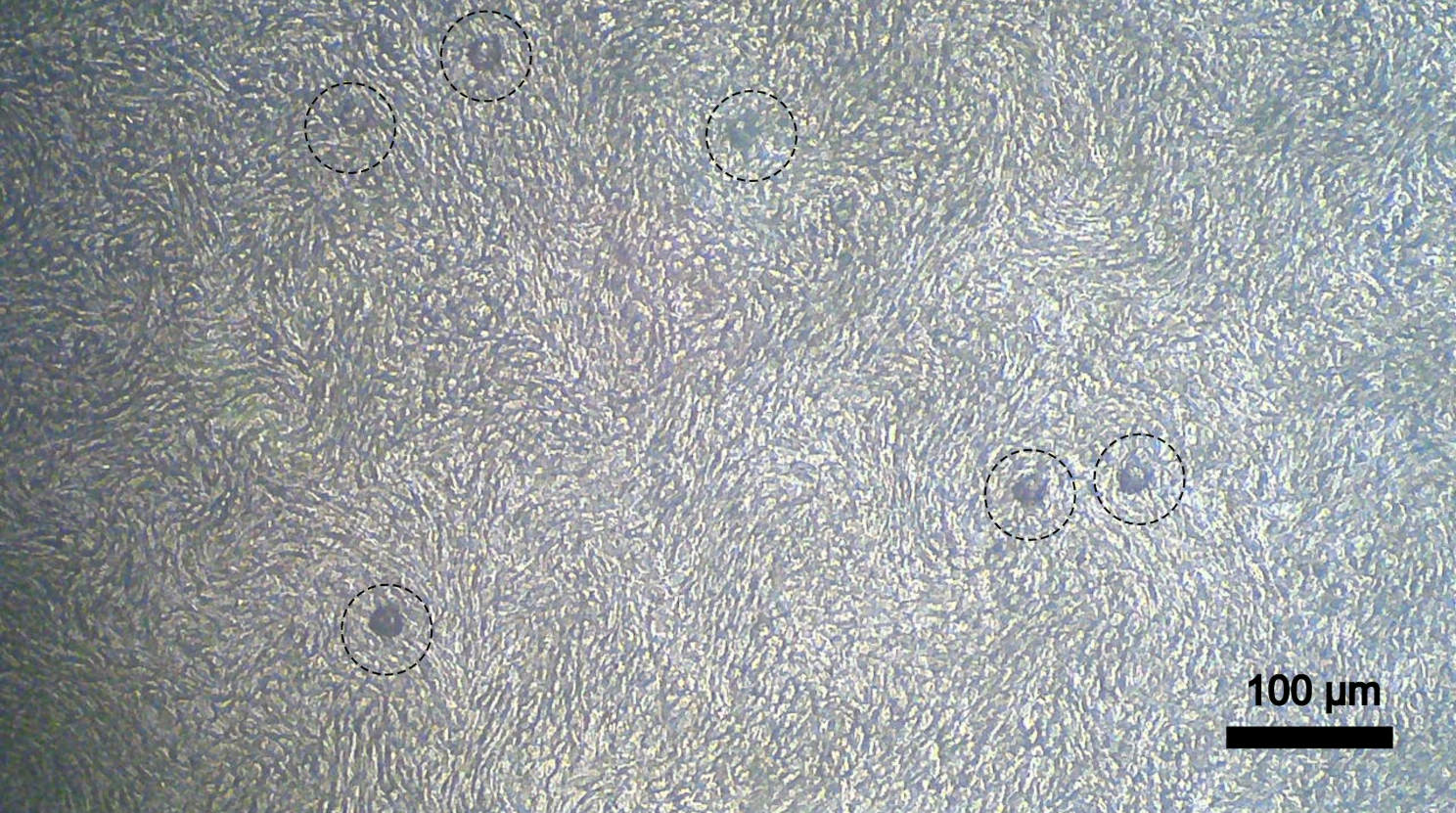
Sensors sit on the surface of airway epithelial cells. The epithelial cells form a Van Gogh-style pattern when grown in the lab.
Not so fast
This proof-of-concept research showed that such sensors could respond to the activity of the ‘healthy state’ of the key protein that causes CF. But more research is needed if we are to understand how the sensors respond to different types of CF-causing mutations, of which there are more than 2000. We also need to know how effectively they could be employed to detect protein function restoration following screening for personalised medicine.
Why does it matter?
With the development of ever more effective, but often expensive life-long treatments for CF, it’s important to know which treatments restore healthy airway chemistry. Testing is done on an individual basis on a patient’s cells because of the huge variation in disease-causing mutations and the hope is that these sensors will provide a meaningful new tool for this screening process.
Take home messages
1. CF requires a personalised approach to therapy.
2. Testing the effectiveness of drugs on personalised airway models grown in the lab can help to determine optimum treatment.
3. Micro-scale sensors made from polystyrene and gold could aid the therapy screening process by reporting on chemical changes at the airway surface.
Guest author: Will Skinner, PhD
This article was written as part of a series of ‘journal club’ summaries for Scientific Writers Ltd., and is based on the following publication:
Title: SERS Microsensors for the Study of pH Regulation in Cystic Fibrosis Patient-Derived Airway Cultures
First Author: Skinner W , et al.
Journal: ACS Sensors.
Date online: 25 April 2024
Other references:
Elborn JS. Cystic fibrosis. Lancet. 2016;388(10059):2519-2531.